
In the medical field, the utilization of cervical spine X-rays plays a vital role in diagnosing various conditions related to the neck and upper back region. Understanding the importance of c-spine X-rays and recognizing the indications for their use are fundamental aspects of medical care.
Importance of C-Spine X-Rays
Cervical spine X-rays, commonly referred to as c-spine X-rays, are essential diagnostic tools that aid healthcare providers in identifying the underlying causes of shoulder, neck, arm or upper back pain. These imaging studies can also reveal issues such as tingling, numbness, or weakness in the arm or hand. One of the primary purposes of c-spine X-rays is to detect fractures in the cervical vertebrae or to assess any dislocations present in the joints between the vertebrae, especially in urgent care settings where quick and accurate diagnosis is crucial.
Indications for C-Spine X-Rays
The decision to perform cervical spine radiographs is based on a range of clinical scenarios, including trauma, infection, atypical pain, limb pain, osteoporosis, and degenerative changes. In cases where computed tomography (CT) is unavailable, it is recommended to conduct a comprehensive set of c-spine views to ensure a thorough assessment.

Particularly, the cervical spine series typically consists of these essential views and projections to provide a comprehensive evaluation of the cervical spine area. While CT scans have become more commonplace for trauma evaluations, the c-spine series remains invaluable, especially for radiographers.
Conducting c-spine X-rays, a lateral projection coupled with an open mouth view is considered fairly sensitive in detecting c-spine fractures, with a low risk of missing significant fractures. The sensitivity can further increase to nearly 100% with the addition of the anteroposterior (AP) projection.
Understanding the significance of c-spine X-rays and recognizing the diverse indications for their usage, healthcare professionals can effectively leverage these imaging techniques to diagnose and manage a spectrum of cervical spine conditions.
Types of C-Spine X-Ray Views
Examining the cervical spine, various imaging views are utilized to provide a comprehensive assessment of potential injuries and abnormalities. Understanding the different types of c-spine x-ray views is crucial in clinical practice to ensure accurate diagnosis and effective treatment.
Common C-Spine X-Ray Views
In routine clinical practice, several common c-spine x-ray views are performed to evaluate the cervical spine thoroughly. These views include:
- Anteroposterior (AP) – Provides a front-to-back view, allowing for the assessment of spinal alignment and structures in the cervical spine.
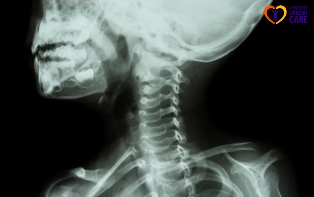
- Lateral – Captures a side view of the cervical spine, highlighting the vertebral bodies, intervertebral discs, and alignment of the spine.
- Obliques – Angled views that help visualize the vertebral foramina and articular facets from different perspectives.
- Odontoid – Specifically targets the dens (odontoid process) of the axis vertebra (C2), essential for detecting fractures or abnormalities in this region.
These common c-spine x-ray views are essential in detecting fractures, dislocations, and other cervical spine injuries. They provide a comprehensive overview of the cervical spine anatomy and aid in the accurate diagnosis of various pathologies.
Specialized C-Spine X-Ray Projections
In addition to the standard views, specialized c-spine x-ray projections offer a more detailed assessment of the cervical spine, especially in complex cases or specific injuries. Some specialized projections include:
- Cervicothoracic View (Swimmer’s View) – Useful for visualizing the lower cervical spine and upper thoracic spine, particularly in cases of trauma or suspected injuries in this region.
- Flexion-Extension Lateral – Assesses spinal stability by capturing the cervical spine in various degrees of flexion and extension. This view helps evaluate motion and potential instability in the spine.
- Fuchs View – A specialized projection that should not be used in a trauma setting due to potential risks. It is used for specific diagnostic purposes in non-urgent cases where trauma is ruled out.
These specialized c-spine x-ray projections provide additional information beyond the standard views, offering clinicians a more in-depth understanding of the cervical spine anatomy and pathology. When utilized appropriately, these views can enhance diagnostic accuracy and guide appropriate management strategies for patients with cervical spine issues.

Reading C-Spine X-Rays
Interpreting C-Spine X-Rays, healthcare professionals employ a systematic approach for accurate analysis. This section covers the interpretation of C-Spine X-Rays and the systematic approach used to analyze these crucial diagnostic images.
Interpretation of C-Spine X-Rays
Radiologists and medical practitioners assess C-Spine X-Rays to identify any abnormalities or injuries in the cervical spine region. A thorough examination involves evaluating the alignment of the vertebrae, the integrity of the bony structures, and the presence of any soft tissue swelling or hemorrhage. Specific areas of focus include:
- Vertebral Alignment: Checking for proper alignment of the cervical vertebrae and assessing for any subluxations or dislocations.
- Bony Structures: Examining the bone density, integrity, and continuity to detect fractures, defects, or abnormalities.
- Soft Tissue: Observing the prevertebral soft tissue width to determine if there are signs of acute swelling or hemorrhage related to an injury.
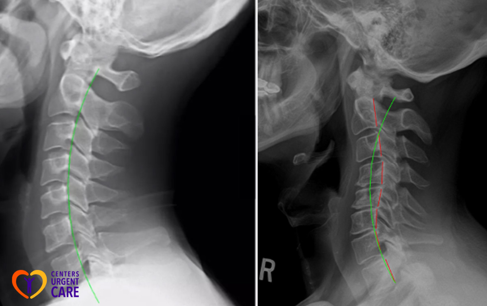
- Spinal Curvature: Assessing the curvature of the cervical spine, including the normal lordotic curve, to identify any deviations.
Systematically analyzing these key components of a C-Spine X-Ray, healthcare providers can accurately diagnose and manage cervical spine conditions.
Systematic Approach to Analyzing C-Spine X-Rays
To reduce the likelihood of missing critical injuries, healthcare professionals adopt a systematic approach when reading cervical radiographs. The process involves visualizing the spine from the base of the skull to the C7-Th1 junction, ensuring comprehensive coverage of the cervical vertebrae. Some specific steps in the systematic approach include:
- Check for Normal Lordotic Curve: Ensuring the presence of a normal smooth lordotic curve in the cervical spine.
- Assess Spino-laminar Line: Verifying the alignment of the spino-laminar line in relation to the vertebral bodies.
- Evaluate Predental Space: Examining the predental space for any abnormalities or signs of injury.
- Assess Prevertebral Soft Tissue: Measuring the width of the prevertebral soft tissues to detect acute swelling or hemorrhage.
- C1-C4 – < 7
- Below C5 – < 22
Employing a systematic approach enhances the accuracy of readings and ensures comprehensive assessment of the cervical spine, aiding in the timely diagnosis and appropriate management of C-Spine conditions.
Identifying Injuries on C-Spine X-Rays
Using c-spine X-rays, healthcare professionals diagnose fractures and dislocations, as well as assess soft tissue injuries in patients. These x-ray images play a crucial role in determining the root cause of various symptoms related to the neck, shoulders, upper back, arms, and hands. Let’s explore how c-spine X-rays aid in diagnosing specific injuries:
Diagnosing Fractures and Dislocations
Cervical spine X-rays are instrumental in pinpointing the underlying issues related to neck and upper body discomfort. They can reveal fractures in the cervical vertebrae or dislocations in the joints between the vertebrae. For instance, when a lateral view of the cervical spine is captured along with an open mouth view, the sensitivity of detecting c-spine fractures is quite high. The addition of an anteroposterior projection further enhances the sensitivity to nearly 100%. This technique allows medical professionals to recognize fractures with high accuracy and minimize the risk of missing significant injuries to less than 1%.
Moreover, emergency physicians utilize c-spine X-rays to diagnose subluxations and dislocations of facet joints by examining the cartilage space between vertebrae corpora, facet joints, and spinous processes. An increased interspinous distance of more than 50% indicates a potential ligamentous injury, aiding in the appropriate treatment and management of the patient.
Assessing Soft Tissue Injuries
In addition to fractures and dislocations, c-spine X-rays are also valuable in assessing soft tissue injuries. The prevertebral soft tissues visible in the x-ray images provide insights into acute swelling or hemorrhage resulting from an injury. Normal measurements of prevertebral tissue width typically decrease from C1 to C4 and increase below C4. Healthcare professionals reference these measurements, with widths of less than 7 mm from C1 to C4 and less than 22 mm below C5 considered within the normal range. By evaluating soft tissue structures on c-spine X-rays, medical practitioners can gauge the extent of soft tissue injuries and tailor treatment plans accordingly.
In summary, c-spine X-rays are pivotal in diagnosing a range of injuries, including fractures, dislocations, and soft tissue damage, enabling healthcare providers to deliver accurate and timely care to patients experiencing neck and upper body complications.
Considerations for Specific Populations
Conducting Cervical-Spine (C-Spine) X-rays, special considerations must be taken into account for specific populations, especially pediatric patients and instances of Spinal Cord Injuries without Radiographic Abnormality.
Pediatric Patients and C-Spine X-Rays
Pediatric patients present a unique challenge when it comes to interpreting C-Spine X-rays. In approximately 25% of pediatric patients with spinal cord injuries, traditional imaging studies such as plain radiographs may not reveal any abnormalities. Therefore, diagnosing potential spinal cord injuries in pediatric patients often requires a comprehensive approach that includes assessing tenderness of the neck and conducting a thorough neurologic examination.
In cases where spinal cord injuries are suspected in pediatric patients, healthcare professionals should not solely rely on imaging results. Instead, a combination of clinical symptoms, physical examination findings, and patient history should guide the diagnosis and treatment plan.
Special Cases: Spinal Cord Injuries without Radiographic Abnormality
In specific scenarios where spinal cord injuries are present without any evident abnormality in radiographic images, known as Spinal Cord Injury Without Radiographic Abnormality (SCIWoRA), a cautious and systematic approach is essential for accurate diagnosis. As mentioned earlier, around 25% of pediatric patients fall into this category, emphasizing the importance of considering alternative diagnostic methods beyond traditional imaging.
Healthcare practitioners managing cases of SCIWoRA must prioritize detailed clinical assessments, including a careful examination of symptoms, patient history, and any subtle neurological signs. In these instances, a high level of clinical suspicion coupled with close monitoring and follow-up care is paramount to ensure appropriate management and positive patient outcomes.
Understanding the nuances of conducting C-Spine X-rays in specific populations, including pediatric patients and cases of Spinal Cord Injuries without Radiographic Abnormality, allows healthcare professionals to deliver comprehensive and tailored care. By addressing the unique diagnostic challenges these individuals present, we can ensure accurate assessments and effective treatment plans. At Centers Urgent Care, we offer fast, high-quality urgent care services in New York City for children and adults. If you’re in need of exceptional care, especially for C-Spine evaluations, don’t hesitate to contact us today. We’re here to provide urgent care that you can trust!
Sources:
2025 has changed my life. I never believed I would be cured by a spell caster who specializes in herbal cure and magical spells. I am talking about the help Doctor Odunga gave to me. I have been cured of herpes HPV which has plagued me for over 2 years. I am very happy to tell others that if you want to be cured or get your ex back spells and get pregnant naturally, even financial blessing through lottery, there is only one place to be and that is with Dr Odunga Spell Temple. Am so happy that just 3 days after taking DR ODUNGA Herbal treatments my herpes was cured permanently. DR ODUNGA also have cure to #HIV #Diabetes #Lupus #Tinnitus #Fibroid #Cancer #Hepatitis B, #Syphilis #Infertility etc … If you have this ailment or whatsoever you might be suffering from you should contact him at his EMAIL: odungaspelltemple@gmail.com OR VIA Whats App +2348167159012