

In today’s healthcare landscape, X-rays have become essential tools, providing invaluable insights into our internal structures, often revealing information that physical exams alone cannot capture. For both healthcare providers and patients, understanding the different types of X-ray imaging available is crucial to recognizing how each type can be applied to accurately diagnose and treat a wide range of health conditions.
Importance of X-Rays in Diagnostic Imaging
X-ray imaging is a cornerstone of diagnostic medicine. It enables healthcare professionals to quickly and accurately view and assess bones, organs, and tissues, which supports faster diagnosis and more effective treatment planning. One significant advantage of X-rays is their ability to produce detailed images non-invasively.
Additionally, X-rays are generally cost-effective, making them accessible for a broad range of diagnostic needs. However, not all X-ray techniques are the same; different types serve specific purposes. Below is a closer look at the most common types of X-ray imaging, each with its applications, benefits, and limitations.

Conventional Radiography: The Basics of X-Ray Imaging
Conventional radiography, or plain film radiography, is one of the most widely recognized and frequently used X-ray techniques. It provides a simple yet effective way to view the internal structures of the body. Here’s a look at how it works and when it is most useful.
How Conventional Radiography Works
In traditional radiography, X-ray beams go through the body and absorbed in varying quantities by numerous kinds of tissue. Dense structures, such as bones, take in more X-rays, resulting in a white image, whereas softer tissues show up in shades of gray. This contrast enables doctors to evaluate bone fractures, look for organ abnormalities, and identify conditions such as pneumonia or osteoarthritis.
Common Applications and Limitations
Conventional X-rays are typically used to:
- Identify Bone Fractures: X-rays clearly show breaks or cracks in bones, helping doctors assess the severity and alignment of fractures.
- Evaluate Dental Health: Dentists use X-rays to detect tooth decay, infections, and bone loss.
- Examine the Chest: Chest X-rays can help diagnose lung diseases, heart conditions, and other thoracic abnormalities.
- Assess Joint Conditions: X-rays can identify issues like arthritis and joint abnormalities.
Though invaluable, conventional radiography does have some limitations. For instance, it may not capture fine details of soft tissues, and repeated exposure to even low-dose radiation may pose risks. Additionally, since conventional X-rays produce two-dimensional images, they may not fully reveal complex anatomical structures.

Computed Tomography (CT): Capturing Cross-Sectional Images
Computed Tomography, or CT, represents an advanced form of X-ray imaging, providing highly detailed cross-sectional views of the body. This technology has become integral in diagnosing numerous medical conditions, especially in emergency and oncology settings.
How CT Scans Work
CT scans use a rotating X-ray device that captures multiple images from various angles, creating a detailed cross-sectional “slice” of the area being examined. These slices can then be combined into a comprehensive 3D image, offering an in-depth view of complex structures and making it easier to identify specific areas of concern.
Advantages and Applications
CT scans provide several notable advantages over standard X-rays, including:
- Enhanced Detail and Resolution: CT scans deliver highly detailed images, particularly useful for visualizing complex body areas.
- Speed and Efficiency: CT imaging is quick, which is beneficial in emergency situations where immediate results are needed.
- 3D Reconstruction Capabilities: CT technology can generate 3D models, helping in detailed assessment and surgical planning.
Applications of CT scans include:
- Trauma Evaluation: CT is invaluable for assessing internal injuries after accidents or other traumas.
- Cancer Diagnosis and Staging: CT scans help in detecting and staging cancers by providing clear images of tumors.
- Guiding Procedures: CT scans are often used to guide biopsies and other minimally invasive procedures.
- Monitoring Treatment: They allow doctors to track the progress of treatments, such as chemotherapy, by measuring changes in tumor size.

Fluoroscopy: Real-Time Imaging for Dynamic Assessment
Fluoroscopy offers unique, real-time imaging capabilities that allow medical professionals to observe the movement of internal structures. This makes it particularly useful for examining dynamic processes within the body.
How Fluoroscopy Works
Fluoroscopy involves a continuous X-ray beam directed through the body, which is captured and displayed as a live video. This real-time capability makes fluoroscopy ideal for procedures where observing movement is essential, such as monitoring the flow of blood or tracking the movement of a swallowed substance through the digestive tract.
Uses in Real-Time Imaging
Fluoroscopy has a wide range of applications across multiple medical fields, including:
- Orthopedics: Doctors use fluoroscopy to visualize joint movements and guide treatments like joint injections.
- Cardiology: This technique assists in procedures like cardiac catheterization, allowing doctors to see the heart in action.
- Gastroenterology: In procedures such as barium swallows, fluoroscopy provides a real-time view of the digestive system.
- Pulmonology: Fluoroscopy assists in bronchoscopy procedures, offering live images of the airways and lungs.
By offering dynamic imaging, fluoroscopy enables doctors to make more accurate assessments, particularly during diagnostic and interventional procedures.

Mammography: Early Detection of Breast Cancer
Mammography is a specialized form of X-ray imaging dedicated to examining breast tissue. This method plays a critical role in the early detection of breast cancer, allowing for timely intervention.
Importance of Mammography
Mammograms can reveal abnormalities in breast tissue long before they are physically noticeable, significantly improving the chances of successful treatment. For women at average risk, annual mammograms are recommended starting at age 40, with some guidelines suggesting every two years after age 55. Women at higher risk may require earlier and more frequent screenings.
Types of Mammography
There are two primary forms of mammography:
- 2D Mammography: This conventional form captures two-dimensional images and is effective for basic screenings, though it may sometimes lead to overlapping images that reduce clarity.
- 3D Mammography (Tomosynthesis): This advanced technique creates a series of cross-sectional images, providing greater clarity and reducing false-positive results, which are common in dense breast tissue.
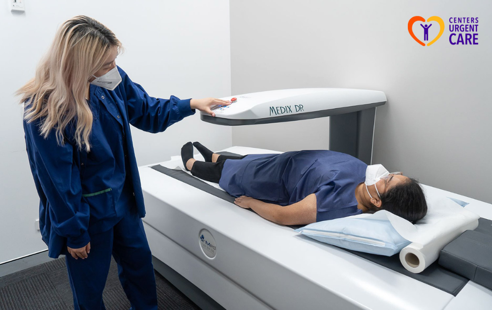
Bone Densitometry: Assessing Bone Health
Bone densitometry, also known as Dual-Energy X-ray Absorptiometry (DEXA), is a specialized form of X-ray imaging used to assess bone density and identify conditions like osteoporosis.
Overview of Bone Densitometry
During a DEXA scan, low-dose X-rays measure bone density in areas such as the spine, hips, and wrists. This data helps determine bone strength and the risk of fractures. DEXA scans are particularly useful for older adults and individuals with risk factors for bone loss.
Uses in Assessing Bone Health
Bone densitometry is critical in diagnosing osteoporosis, tracking bone density changes over time, and evaluating the effectiveness of treatments for osteoporosis. For patients at high risk of fractures, regular bone density assessments can lead to preventive measures that strengthen bone health.
Empowering Health Through Advanced X-Ray Imaging
From the versatility of conventional radiography to the high-resolution insights of CT scans, the real-time benefits of fluoroscopy, and the early detection capabilities of mammography and bone densitometry, each imaging technique offers unique advantages that are crucial for a range of medical needs.
Understanding these various types of X-ray technologies not only empowers healthcare professionals to make more precise diagnostic decisions but also allows patients to take an active role in their health journey. When patients and providers are informed about the purpose and benefits of these imaging tools, they can work together more effectively to address health concerns and prioritize well-being.
At Centers Urgent Care, we are committed to offering reliable, high-quality care in New York. Our experienced team prioritizes patient well-being and provides prompt, thorough services you can trust. Discover why we’re recognized as the best urgent care in New York. Contact us today to learn more about how we can support your health!
2025 changed my life. I never believed I would be cured by a spell caster who specializes in herbal cure and magical spells. I am talking about the help Doctor Odunga gave to me. I have been cured of herpes HPV which has plagued me for over 2 years. I am very happy to tell others that if you want to be cured or get your ex back spells and get pregnant naturally, even financial blessing through lottery, there is only one place to be and that is with Dr Odunga Spell Temple. Am so happy that just 3 days after taking DR ODUNGA Herbal treatments my herpes was cured permanently. DR ODUNGA also have cure to #HIV #Diabetes #Lupus #Tinnitus #Fibroid #Cancer #Hepatitis B, #Syphilis #Infertility etc … If you have this ailment or whatsoever you might be suffering from you should contact him at his EMAIL: odungaspelltemple@gmail.com OR VIA Whats App +2348167159012